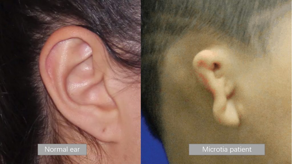
Microtia is a congenital deformity where the auricle (external ear) is underdeveloped. Traditional treatment often entails suturing multiple pieces of rib cartilage to form the ear, but this is a challenging surgical because everyone is unique and the shape of the ear varies so much.
It is challenging for the reconstructed ear to reproduce all the complex and subtle 3D structural features of the natural ear. Guigang People’s Hospital’s Burn and Plastic Surgery Department has introduced high-precision 3D digitalization technology to improve bilateral symmetry effect after auricular reconstruction, using an objective and accurate digital reconstruction ear positioning method and special stent design method.
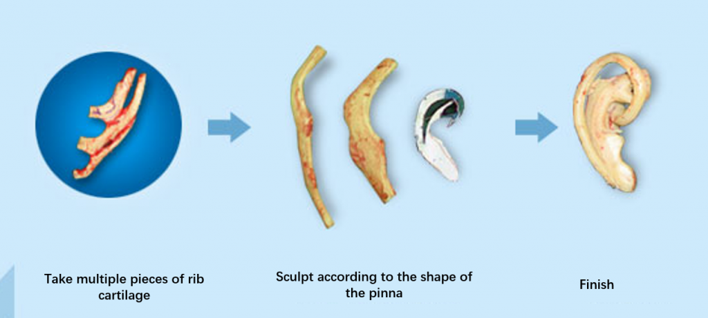
Table of Contents
Challenges
· Complex ear stucture
There are many fine structures and it is difficult to carve. It requires the operator to have not only plastic surgery repair and reconstruction skills, but also superb carving skills and high aesthetic level.
Different types of microtia
· Difficulty in selecting a reference object
Generally speaking, the natural ear on the normal side is the perfect reference object for reconstructing the shape of the ear in patients with microtia. However, even when a reference object is available, the doctor still needs to translate the ear in three dimensions in the brain, and imagining the reconstruction of the shape of the other side of the ear requires a very high level of 3D imagination on the part of the doctor.
3D Digital Solution
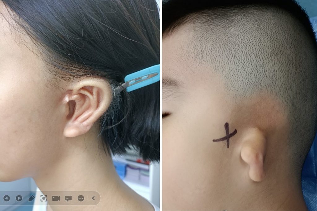
Therefore, the doctors in the Burn and Plastic Surgery Department of Guigang People’s Hospital used the Shining 3D EinScan Pro HD multifunctional handheld 3D scanner to obtain the 3D data of the patient’s natural ear on the normal side non-contact and quickly.
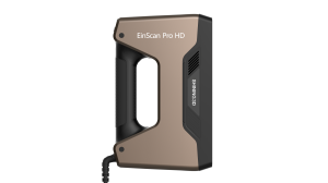
EinScan Pro HD
The highest accuracy of EinScan Pro HD scanner can reach up to 0.04mm, and the minimum point distance can reach 0.2mm, which can restore the 3D shape of the patient’s normal ear in high detail and post-processed in a clean STL model. The whole procedure was finished in minutes.
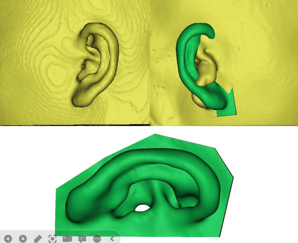
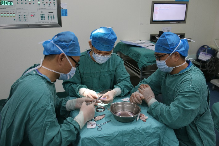
Digital model and the reconstruction process
Based on the high-precision 3D model of the patient’s healthy ear collected by the 3D scanner, researchers from the Guangxi Digital Medicine and 3D Printing Clinical Medical Research Center reconstructed the 3D data of the healthy ear by smoonthing, filling defective areas to a closed and clean mesh, and this mesh was then mirrored as the 3D structure of the ear on the affected side to be reconstructed.
What’s next
The researchers then imported the 3D design data of the ear into a 3D printer to customize the patient’s ear reconstruction template, which was then sterilized and used in the operating room by plastic surgeons.
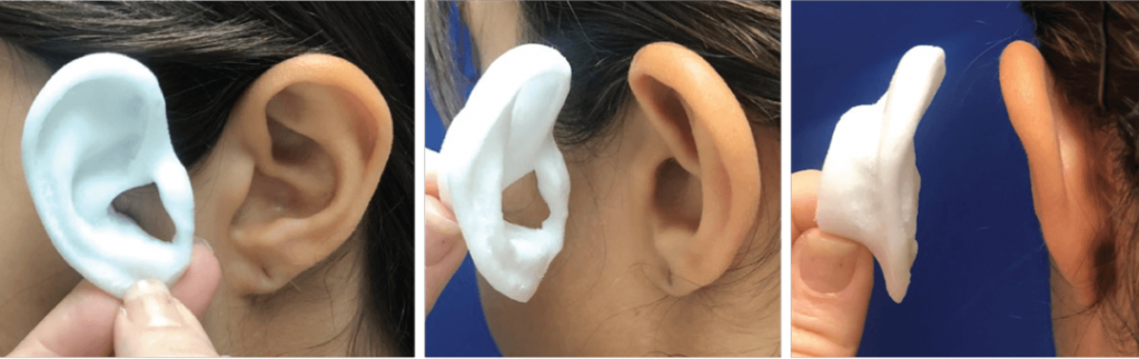
Based on the 3D printed template of the reconstructed ear, the surgeon can hold and analyze the shape and relief closely, which provides a great start to creat a cartilage sculpture in the correct size and shape.The method of digital process of using 3D scan to print helped saving surgical time and the amount of cartilage in use compared to the tranditional way.
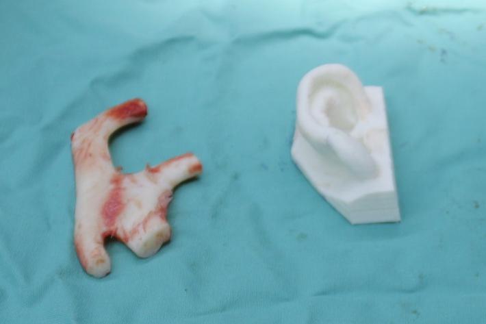
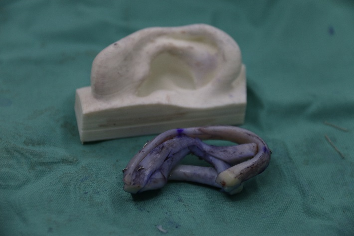
The finished cartilage sculpture
With almost one in 6,000 children suffering from congenital microtia, children all over the world can change their lives with this surgery. With the assistance of the EinScan Pro HD 3D scanner, doctors are bringing hope to one child after another.
The material in this article belongs to the original author, if there is any infringement, please contact to delete.


