In January 2022, Prof Matthew Brookes, a physicist from the University of Nottingham, won a physical science and engineering prize for helping to invent a new wearable brain scanner.
Researchers say it can revolutionize brain imaging by identifying the parts of the brain responsible for activities such as nodding, drinking tea, and playing with bats and balls. The system is currently being installed in a clinical setting, in collaboration with the national charity Young Epilepsy and Sick Kids in Toronto.
The introduction of Prof. Matthew Brookes’ Project by Nature
Professor Brooks said the wearable scanner has been in development for six years. The EinScan H 3D scanner is an important member of their research into brain activity.
Traditional magnetoencephalogram (MEG) scanners tend to be large and very bulky, and suffer from the one-size-fits-all problem. In order to study brain activity during different activities, participants need to perform several movements, so wearing a helmet equipped with an OPM sensor becomes a more feasible option.
There are two ways to firmly mount the OPM sensor on the scalp. The first is to use a subject-specific helmet based on their anatomical MRI scan. This allows for accurate knowledge of the position and orientation of the sensor relative to the participant’s head, but is expensive and requires MRI scans for all participants, which is a huge challenge for commercialization.
 A 49-channel whole head brain scanner.
Image Credit: University of Nottingham.
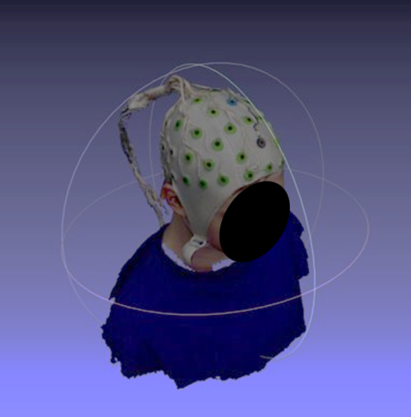
A second option is to use a generic helmet that can accommodate a larger portion of the population. However, the position of the sensors in each person’s head would be different and therefor Professor Matthew Brooks used the EinScan H handheld 3D scanner to help customize the 3D printed helmet and place sensors onto the helmet to reconstruct brain activity. They found that the EinScan H was able to quickly generate a digital twin of the participant and co-align the OPM-MEG sensor position to the participant’s MRI to reconstruct brain activity.

EinScan H is a hybrid light source of LED and infrared. Scanning data accuracy of up to 0.05 mm and volume accuracy of 0.1 mm/m improves the accuracy of 3D modeling in dense point clouds or polygon meshes and helps to accurately locate the head sensor. At the same time, the face scanning method uses invisible infrared light for a safe and comfortable scanning process, which is useful for children and people with photosensitive epilepsy.

“Our group in Nottingham, alongside partners at UCL, are now driving this research forward, not only to develop a new understanding of brain function, but also to commercialize the equipment that we have developed.” Brooks concluded, and thanks to EinScan H 3D scanner, this blueprint is gradually becoming possible.
If you are as fascinated as we are about the work the team at Nottingham University is doing, watch the video: https://youtu.be/9FrsYjkKhBI
This case originated from: https://www.3dscantocad.co.uk/post/nottingham-university-and-einscan-h-3d-scanning-in-healthcare
Reference: Hill, R. M., et al. (2020) Multi-Channel Whole-Head OPM-MEG: Helmet Design and a Comparison with a Conventional System. NeuroImage. doi.org/10.1016/j.neuroimage.2020.116995.


